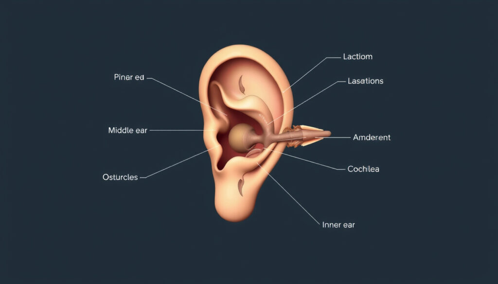
Parts of the Ear: Anatomy, Diagram & Functions
You use your ears every waking moment, yet most people couldn’t name more than one or two parts. That changes now. Your ear has three main sections—outer, middle, and inner—that work together as a team, and understanding how they fit is surprisingly straightforward once someone shows you the map.
Main parts: 3 (outer, middle, inner) · Bones in middle ear: 3 ossicles (malleus, incus, stapes) · Outer ear components: pinna, ear canal · Middle ear includes: eardrum, ossicles · Inner ear key parts: cochlea, semicircular canals
Quick snapshot
- The ear has three main sections: outer, middle, and inner (Cleveland Clinic)
- Ossicles are three tiny bones: malleus, incus, stapes (Cleveland Clinic)
- Some sources mention “six bones” in the ear—likely a miscount combining ossicles with ear canal components
- Exact ossicle dimensions rarely appear in consumer-facing materials
- Ear anatomy knowledge has been consistent—no recent structural discoveries
- Modern imaging has refined understanding of cochlear hair cells
- The article walks through each section with names, locations, and jobs
- Kid-friendly terminology included throughout
Ear anatomy follows a clean three-part architecture: one section collects sound, one amplifies it, and one converts it to signals the brain understands.
The three main parts of your ear include the outer ear, middle ear and inner ear. The outer ear includes the pinna, temporal bone, and ear canal.
The ears job is to convert the sound energy into a neutral signal which later is received by your brain.
| Structure | Value |
|---|---|
| Main sections | Outer, middle, inner |
| Ossicles count | 3 |
| Hammer bone | Malleus |
| Anvil bone | Incus |
| Stirrup bone | Stapes |
What are the sections of the ear?
Every ear you meet belongs to one of three neighborhoods. The Cleveland Clinic puts it plainly: the outer ear, middle ear, and inner ear. Each has a distinct job and a distinct environment—air on the outside, air in the middle cavity, fluid in the inner sanctum.
The outer ear is the only part you can see and touch. The middle ear is hidden behind the eardrum but still air-filled. The inner ear sits deep in the skull, wrapped in bone, and filled with fluid that does the actual translating.
Outer ear
The outer ear gathers sound waves and funnels them inward. Think of it as the satellite dish of the hearing system—always pointed at the world.
Middle ear
The middle ear takes vibrations from the eardrum and cranks up the volume before passing them along. Three tiny bones handle that amplification job. It’s a loud space compared to the quiet inner ear.
Inner ear
The inner ear is where hearing actually happens. Fluid fills the cochlea, tiny hairs catch the wave motion, and electrical signals shoot to the brain via the cochlear nerve. The same section runs balance through the semicircular canals.
The pattern across all three is simple: collect, amplify, convert. Each section does one thing and passes the job to the next.
The stapes (say: STAY-peez) (“stirrup”), which is attached to the incus and is the smallest bone in the body.
What are parts of the ear called?
The names matter because they unlock the diagrams. Once you know malleus means hammer, the shape makes perfect sense. The NIDCD illustration of ear parts labels each piece clearly, and matching those labels to real structures is half the battle.
- Pinna (also called the auricle)—the visible flap
- Malleus (hammer)—attaches directly to the eardrum
- Incus (anvil)—bridges the hammer and stirrup
- Stapes (stirrup)—the smallest bone in the human body
The Latin names for the ossicles translate directly to their shapes. Malleus means hammer, incus means anvil, stapes means stirrup. That visual memory trick makes the names stick.
Pinna and ear canal
The pinna catches sound waves and directs them down a narrow tube called the external acoustic meatus (or ear canal for short). That canal runs about 2.5cm long before hitting the eardrum.
Eardrum and ossicles
The tympanic membrane (eardrum) is a tight sheet of tissue that vibrates when sound hits it. Those vibrations shake the malleus, which shakes the incus, which shakes the stapes. Each bone amplifies the motion like a tiny lever system.
Cochlea and vestibule
Once the stapes passes vibrations to the inner ear, the cochlea takes over. The Cleveland Clinic describes it as a snail-shaped organ filled with fluid. Tiny hairs lining the cochlea bend with each wave and send electrical signals to the brain. The vestibule sits nearby and handles balance detection alongside the semicircular canals.
The implication: the outer ear handles collection and direction, the middle ear handles amplification and pressure, and the inner ear handles translation into signals the brain understands.
What are the parts of the outer ear?
The outer ear has two main pieces: the pinna you can see and the ear canal that carries sound inward. Nemours KidsHealth explains that the pinna is made of cartilage covered by skin—the firm but flexible stuff you can bend without breaking.
Auricle (pinna)
The auricle has ridges and valleys that help directional hearing. The McGovern Medical School notes the specific parts: tragus (the little bump in front), helix (the outer rim), and lobule (the earlobe). Cartilage gives the pinna its shape; skin covers and protects it.
External acoustic meatus
The ear canal is roughly an inch long, lined with skin that produces cerumen (ear wax). That wax traps dirt and dust before they reach the eardrum. Glands in the canal lining also provide a bit of waterproofing against moisture.
The implication: the outer ear does more than collect sound. It filters and directs it, getting sound waves ready for the eardrum with a little help from ear wax along the way.
What is the tiny bone in the middle ear shaped like a hammer?
The malleus. Its Latin name translates directly to “hammer,” and if you’ve ever seen one, the comparison clicks immediately. The malleus sits right next to the eardrum and vibrates with it whenever sound waves hit.
Malleus
The malleus is the first of three ossicles. It attaches to the inner surface of the eardrum and passes vibrations to the incus. Its handle (manubrium) grips the eardrum; its head connects to the incus.
Incus and stapes
The incus is the middle piece—shaped like an anvil, hence the name. It receives vibrations from the malleus and passes them to the stapes. The stapes is the final bone, shaped like a stirrup, and its footplate fits into the oval window of the cochlea.
According to Nemours KidsHealth, the stapes is the smallest bone in the human body. It measures only about 3mm—smaller than a matchstick head.
These three bones are the only ones in the body specifically designed for hearing. They work as a mechanical amplifier, boosting sound energy roughly 20 times over before passing it to the fluid-filled cochlea.
What are parts of the ear and their functions?
Each section has a clear job. The Cleveland Clinic lists the primary functions as hearing and balance—and everything in ear anatomy serves one of those two missions.
Outer ear functions
- Pinna collects and funnels sound waves toward the ear canal
- Ear canal directs sound to the eardrum while protecting it
- Ear wax traps particles before they reach the eardrum
Middle ear functions
- Eardrum converts sound waves into mechanical vibrations
- Ossicles amplify vibrations and transmit them to the inner ear
- Eustachian tube equalizes air pressure on both sides of the eardrum
Inner ear functions
- Cochlea converts vibrations into electrical signals the brain can read
- Semicircular canals detect head rotation and maintain balance
- Vestibule senses linear motion and head position relative to gravity
What this means: the outer ear handles collection and direction, the middle ear handles amplification and pressure, and the inner ear handles translation into signals the brain understands. They never overlap—they pass the baton.
Related reading: fever guide · cellulitis signs
twinkl.com, istockphoto.com, teacherspayteachers.com, childrenswi.org, freepik.com
While the ear’s intricate anatomy supports hearing and balance, signs of ear infections such as sharp pain or fluid buildup often arise in the outer or middle sections.
Frequently asked questions
What does an ear parts diagram show?
An ear diagram typically shows the pinna, ear canal, eardrum, three ossicles (malleus, incus, stapes), cochlea, semicircular canals, and Eustachian tube. Professional medical posters from ASHA include all these structures labeled clearly.
What are parts of the ear for kids?
Kids learn the big three: outer ear (pinna and canal), middle ear (eardrum and three tiny bones), inner ear (cochlea for hearing, semicircular canals for balance). Nemours KidsHealth explains these in plain language with relatable comparisons.
What is anvil ear?
“Anvil ear” is not a real condition—the phrase is a mishearing of “incus,” the middle ossicle. Incus means anvil in Latin, and some people confuse the two words when hearing about ear anatomy for the first time.
What are the six structures of the inner ear?
No standard list of “six” inner ear structures exists. The key parts are the cochlea, semicircular canals (three of them), vestibule, and the nerves connecting them to the brain. Some sources count individual canal pairs as separate structures, which may explain the confusion.
What are the six bones in the ear called?
There are only three bones in the ear—the ossicles. The “six bones” claim may come from diagrams that label both the ear canal components and the ossicles separately. The actual ossicles are malleus, incus, and stapes.
What are parts of the ear piercing?
Ear piercings pass through skin and sometimes cartilage of the outer ear—the pinna or earlobe. The anatomy of the ear doesn’t change for piercing purposes, but cartilage piercings heal more slowly than lobe piercings because cartilage has less blood flow.
More related posts
 Shoppers Drug Mart Medicine Hat: Hours, Locations & Services
 Chris Noth: Career Fallout from Allegations and Firing
 Royal Challengers vs Kings: IPL Final Scorecard & Highlights
 Geena Davis: Biography, Health Misdiagnosis, and Advocacy
 Bryan Johnson: Age Reversal, Net Worth and Blueprint
 Where Are the Olympics 2026 – Milano Cortina in Italy
 Richard Branson Net Worth, Family & Health Profile
 Mackenzie Gunther: Air Canada First Officer Killed in Crash
